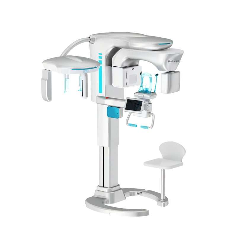
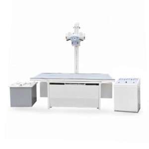
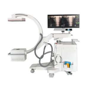
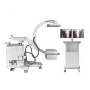
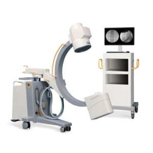
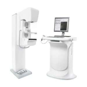
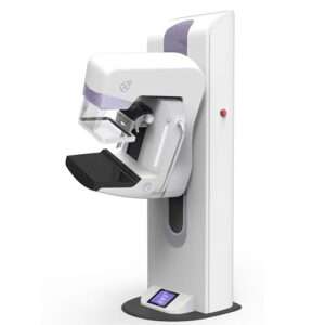
GALAXY DREAM CBCT
GALAXY DREAM CBCT
GALAXY DREAM CBCT
Product Introduction
Model: SHO-3DC780
Features:
- Low radiation: The pulsed exposure dose is 50% less than continuous exposure.
- High voltage generator: 400khz ultra-high inverter frequency, heat dissipation capacity, insulation capacity, improve the stability of the equipment, and be able to continuously shoot 200 groups of CT data.
- Motion Compensation Artifact Algorithm: Effectively removes motion artifacts and can improve image quality by 60%.
- AI low-dose algorithm: At the same time as the low dose, the image algorithm is used to ensure the image quality.
- New 4-in-1: CT, panorama, head and side, integrated sliding seat,can sit and stand to meet the needs of all patients in an all-round way.
- Integrated sliding seat: can be sitting, standing, sliding and rotating dental tablet. Sitting shots are more stable and more comfortable.
- Adaptive head clamp: Head clamp automatic retraction and release
- monitoring: During the shooting exposure, if the patient has a large movement, a pop-up window will prompt, and the monitoring video will be retained for doctor-patient communication.
- Large field of view 18*14: One imaging, able to see the temporomandibular joint and complete maxillary sinus.
- Intelligent post-processing software: is convenient for clinical operation, simple, efficient and smooth. Lateral automatic tracing, bone age analysis, and orthodoxy analysis.
Technical data FOV 18*14 or 16*12 CT/OPG detector
Detector
a-Si Flat Panel Detector
Scintillator
Cesium Iodide
Minimum Voxel Size
0.05mm
Pixel Size
Image pixel: 98μm
Scan Time for CT24S,18S,8S
Freely Adjusting the FOV in CBCT Mode
Yes
Ceph Detector
Detector
a-Si Flat Panel Detector
Scintillator
Cesium Iodide
Pixel Size
Detector Effective Field of View: 233.1mm×7.2mm
Transmission Mode
Wired Transmission
High Voltage Generator
X-ray Generator Power1200W
Working Pattern
Continuous/Pulsed
Tube Voltage60kV~100kV
Tube Current2mA~12mA;
Convert Frequency400kHZ
Exposure Mode
Pulsed/Continuous Exposure
X-ray Tube
Focal Spot0.5mm
Target Angle5°
Tube Voltage60kV~100kV
Tube Current2mA~12mA;
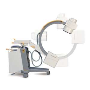
Gantry
Vertical Range of Column Movement690mm
Rotating arm
Rotational bias ≤±1°
Translation Range of Rotational Axis
Translation Distance: 0-86mm, Bias ≤±5mm
Functions
CT/Panoramic Jaw Rest
TMJ Jaw Rest
Temporal Clip
Head Clip
Movable Jaw RestYes
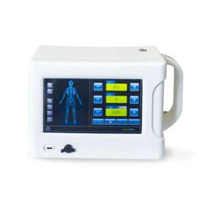
Touch Screen
InstallationInstallation Method: Detachable Mobile Touch Screen
Functions
Rise: Controling the rise of the column
Fall: Controling the fall of the supporting column
Capture Mode: Simultaneously displaying the capture mode
Voltage: Displaying the preset voltage and adjusted voltage during capture
Current: Displaying the preset current and adjusted current during capture
Automatic Ear Clip: Loosening/Tightening the temporal clip
Positioning Laser: Turning on/off the foot positioning laser
Partial Adjustment: Adjusting the height of the jaw rest
Workstation
Operating Systems
Memory:6GB
Disk capacity:500GB
Display Card:High-speed Image Processing Board
CD-ROM: DVD/CD-ROM Drive
Network Card: Gigabit Network Card
Screen Size: 23 inches
Screen Type: LCD, Color
Image Processing Algorithm
Metal Artifact Correction Algorithm
Image Acquisition ModeCT/OPG/CEPH
Auto Focus
Auto Focus(Third-Party Certification provided)
Patient Acquisition
Optional Modes: Children, Elderly, Male, Female
Exposure settings
Tube current, Tube voltage, Voxel size
Image Browsing and Editing
Browsing the uploaed and captured images (Including CT, OPG, CEPH)
Changing the contrast/luminance of images
Image zooming
Dragging the image
Edit list: Text/ Curve/Ellipse/Rectangle/Polygon with arrows, remark
Image Sharpening
Calling the API and passing in the corresponding coefficients to get the adjusted image
Adjusting the window width and window level in the Mater software
Defaulting sharpened image loaded in the background
Customizing the coefficients in the configuration page to change the sharpening effect in real time
Automatically Marking the Neural Tube
With the AI algorithm, the neutral tube could be marked automatically in the panoramic image and also displayed through the image in the MASTER software.
Scanning the QC Code to Get Panoramic Image
Patients can scan the QR codes to download their panoramic images
Enabling the storage and transmission of electronic image


Through professional online marketing platform and offline medical exhibition and worship business modes, the company has a large number of loyal and strong distribution partners, products have been exported to many countries, the company’s Shoimage brand has gained very good influence
Contact Us
Avenida Padre Manuel De Nobrega 11 B
Contribuinte N. 516052632
1000-223 Lisboa